
Research Article
Abstract
Aloe vera (Aloe barbadensis Miller) is orally ingested by many, because of its medicinal benefit in skin treatments, digestive system disorders and improvement of blood sugar among others. Although, Aloe vera has been reported to have different protective effects, no study has investigated its toxicity on the Liver and kidneys at varying concentrations. Therefore, this study aims to evaluate the toxicity potential of Aloe vera on the liver and kidney. Twenty (20) adult albino rats, weighing (151±10 g), were randomly grouped into four groups; A, B, C and D. Group A rats were used as normal controls and received no administration. Group B received a high dose (6 mL /kg) of Aloe vera juice (AVJ). Group C received a medium dose of AVJ (4.5 mL/kg), and group D received a low dose of AVJ (3.0 mL/kg), daily for 21 days. The liver was assessed by estimating serum total bilirubin and liver enzymes (AST, ALT, and ALP) levels. Nephrotoxicity was assessed by measuring Serum creatinine, urea, Na+ and K+. Administration of a high dose AVJ resulted in significantly elevated levels of the liver biochemical parameters and a significant increase in the concentration of BUN, Creatinine, K+ and reduction in Na+ levels. At a medium dose, there were non-significant alterations in the biochemical parameters of the liver and the kidney. At a low dose, there were only slight changes in these biochemical markers. Histopathological studies were in tandem with the biochemical observations. This study reveals that Aloe vera gel possesses a dose-dependent hepatorenal toxicity.
Keywords
Aloe vera, Aloe barbadensis Miller, hepatotoxicity, nephrotoxicity, phytochemicals
1. Introduction
Aloe vera is a desert plant with the botanical name Aloe barbadensis Miller [1]. It is also known as the ‘healing plant’ and the
‘wonder plant’ [2]; it is believed to be very effective in treating
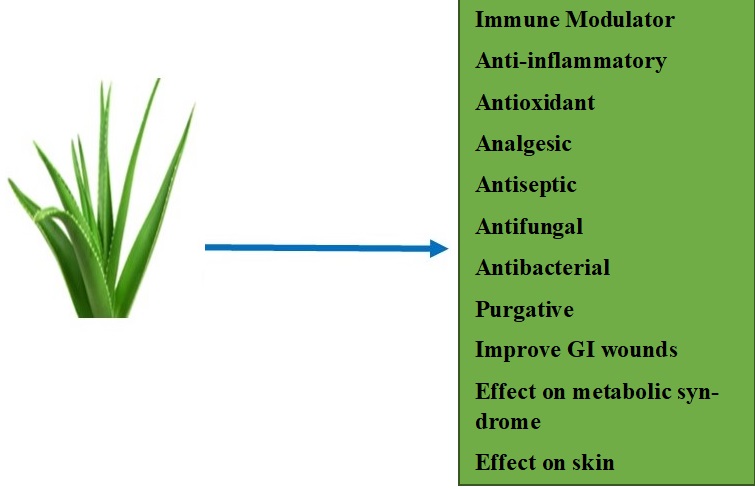
burns and wounds, and has a wide variety of uses (Fig, 1). Aloe vera has
been known to contain several compounds including vitamins, glycoproteins,
minerals, amino acids, phytonutrients, enzymes and glyconutrients with a very
high water content [2]. In addition, it has other pharmacological
activities including anti-inflammatory activities, anti-cancer, anti-diabetic
and antimicrobial activities due to its anthraquinone content [3].

Figure 1. Therapeutic benefits of Aloe vera [4]
Aloe vera helps with intestinal absorption and has anti-ulcer properties. It is an excellent antioxidant and provides metabolic help due to its high vitamin content. When aloe vera nutrients are consumed, they are digested and absorbed into the bloodstream via the intestines, eventually reaching the liver and the kidneys, where they are metabolised and metabolic byproducts eliminated respectively.
The liver's histoarchitecture is designed in such a way that any changes would result in liver injury [5], which could be acute or chronic. Acute liver injury is defined as a sudden decrease in liver cell function in patients without any prior liver illness [6]. Chronic liver illnesses are defined as persistent inflammation, progressive hepatic cell death and regeneration, liver progenitor cell proliferation, and fibrosis. The extracellular matrix is deposited after liver damage to promote wound repair and regeneration; however, recurrent injury from xenobiotics and diseases can lead to progressive fibrotic response characterized by substantial collagen deposition [7]. Hepatocytes have organelles in their cytoplasm that synthesise enzymes; liver injury, whether acute or chronic, alters the function and levels of these enzymes [8]. The kidney plays a very important role in the elimination of wastes from the body. However, when there is decreased blood flow to the kidney, inflammation or excessive exposure to toxins. an acute kidney injury (AKI) develops [9].
Various studies have established the nutritional and medicinal importance of Aloe Vera. However, the potential toxicological side effects of this plant at different concentrations have not been studied in-depth. Therefore, the aim of this study is to investigate the potential hepatorenal toxicity of Aloe vera.
2. Materials and methods
2.1 Plant materials
The fresh Aloe vera (Aloe barbadensis miller) used for the study was procured from Ogbete market, Enugu state, Nigeria. A consulting taxonomist at the Department of Plant Science and Biotechnology's herbarium section, University of Nigeria, Nsukka, verified the authenticity of the plant material
2.2 Reagents
Laboratory kits (Randox, UK) for alanine aminotransferase, aspartate aminotransferase, alkaline phosphatase, and bilirubin were bought from Alpha Pharmaceuticals in Enugu, Nigeria.
2.3 Aloe vera juice (AVJ) extraction
This was prepared according to the preparation by Embark and Abdalla [10], with slight modifications. Fresh Aloe vera plants were washed. The bottom scarred end and thorny sides were cut off to ensure that the gel is carefully separated from the leaves. About 20g of the gel was homogenised in a Qasa blender (Nigeria). It was blended until it was frothy and liquefied. The resultant Aloe vera juice (AVJ) was stored at 4oC in the refrigerator.
2.4. Animals
A total of twenty (20) adult albino rats, weighing 151 ± 10 g, were obtained from the animal house of the College of Veterinary Medicine, University of Nigeria. The animals were housed in metallic cage under standard conditions of temperature (22 ± 3oC) and a 12 h light, 12 h dark cycle. The animals were monitored for 14 days earlier than the experiment's start in order to allow them to acclimatize to the environment. The experimental design and management complied with institutional regulations detailing the use of rats and the guidelines for the care and use of vertebrates in the study published by the American Physiological Society (APS); and the ethical approval for the study was given by the Ethics Committee of Faculty of Veterinary Medicine, University of Nigeria, Nsukka, Nigeria. (Approval number: UNN/eTC/14/69258).
2.5 Phytochemical analysis of the aloe vera
Aloe barbadensis Miller was screened for flavonoids, glycosides, saponins, tannins, steroids, proteins, carbohydrates, and terpenoids by the Department of Pharmacognosy, University of Nigeria Nsukka. Trease and Evans [11] methodology was applied for the analyses.
2.6 Design
The twenty (20) albino rats were grouped into 4 groups (A-D); five rats in each group. The protocol used in the present study is the same as described previously by Embark and Abdalla [10]; who administered 20 mL/kg aloe vera juice to experimental rats (200-250 g weight), once daily for 14 consecutive days. We chose a smaller volume of dosage to ensure safety and effective experimental finding. Bt trial and error we administered graded doses 3 mL/kg, 4.5 mL/kg and 6 mL/kg aloe vera juice to separate groups of rats (150-160 g weight), once daily for 21 consecutive days. These concentrations are far below the oral LD50 of aloe vera juice reported by Jimmy et al [12] to be 1870.83 mg/kg. The administrations were carried out for 21 days as follows:
Group A (Control): No treatment
Group B: received oral administration of a high dose of AVJ (6 mL/kg)
Group C: received oral administration of a medium dose of AVJ (4.5 mL/kg)
Group D: received oral administration of a low dose of AVJ (3 mL/kg)
2.7 Animal sacrifice and sample collection
Under chloroform anesthesia, blood samples for biochemical analysis were taken from the left ventricle of the heart. The liver and kidneys were excised for histological examination.
2.8 Biochemical analysis
2.8.1 Measurement of liver function biomarkers
Reitman and Frankel's colorimetric approach was used to determine Alanine transaminase (ALT) and Aspartate transaminase (AST) [13]. The colorimetric method for assessing ALP activity was developed by Kind and King [14]. Total bilirubin was measured using the colorimetric method published by Malloy and Evelyn [15].
2.8.2 Measurement of renal function biomarkers
Electrolyte, urea, and creatinine levels in the blood were measured to assess renal function. Perlong Medical PL1000A Electrolyte Analyzer was used to measure serum K+ and Na+ level. Urea level was estimated using diacetylmonoxime technique involving protein precipitation [16], while creatinine level was estimated using Jaffe’s method [17].
2.8.3 Histopathological analysis
The paraffin wax embedding method was used to prepare the removed liver and kidney tissues. Sections of each organ were made at a thickness of 5 microns, and Hematoxylin and Eosin staining technique was used for better general examination of the tissues [18]. An OlympusTM light microscope was used to examine the tissue sections.
2.8.4 Statistical analysis
Version 7.0 of Graph Pad Prism (San Diego, CA, USA) was used to analyze the data. The results of the biochemical experiments were presented as mean ± SEM (standard error of the mean). The level of significance was assessed using one-way analysis of variance (ANOVA). Probability levels below 0.05 (p<0.05) were taken as being significant.
3. Results
Table 1 shows the findings of the phytochemical examination of Aloe barbadensis Miller. The results showed that methanol extracts of Aloe barbadensis Miller are strongly rich in saponins, tannins, carbohydrates and flavonoids; glycosides and proteins are present in medium amounts; alkaloids and resins are present in small amount; while reducing sugar, acidic compounds, oils, terpenoids and steroids are absent.
Table 1. Phytochemical analysis of Aloe barbadensis Miller
Compounds | Indication |
Saponins | Strong |
Reducing Sugar | Absent |
Alkaloids | weak |
Tannins | Strong |
Carbohydrates | Strong |
Glycosides | Medium |
Resins | Weak |
Flavonoids | Strong |
Steroids | Absent |
Terpenoids | Absent |
Oils | Absent |
Acidic Compounds | Absent |
Proteins | Medium |
Key: strong= +++; medium=++; weak= +; absent= - according to Trease and Evans [11] | |
3.1 Biochemical results
Liver function was assessed by determining the serum level of the liver biomarkers; ALT, AST, ALP and total bilirubin. A statistically significant (p < 0.05) elevated levels were seen in group B (AVJ 6 mL /kg, high dose) and group C (4.5 mL/kg, medium dose) when compared with groups A (control) and D (3 mL/kg, low dose) (Table 2).
Renal function was assessed by determining the biochemical markers; creatinine, blood urea nitrogen (BUN), potassium (K+), and sodium (Na+) (Table 1). From the results, a statistically significant (P<0.05) elevated levels of blood urea nitrogen (BUN), creatinine and potassium (K+) were seen in group B (AVJ 6 mL/kg, high dose) and group C (AVJ 4.5 mL/kg, medium dose) when compared with group A (control). Furthermore, we observed that the Aloe vera gel (3 mL/kg, low dose) did not pose any renotoxic effects in the animals (Table 3).
Table 2. Statistical comparison of Liver biochemical markers of the experimental animal groups.
Groups | ALT (IU/L) | AST (IU/L) | ALP (IU/L) | Total Bilirubin (mg/dl) |
A: control | 20.67 ± 0.88* | 26.00 ± 1.15** | 236.67 ± 2.85*** | 0.93 ± 0.06** |
B: High dose AVJ (6 mL/kg)) | 27.00 ± 2.08 | 38.00 ± 1.73 | 382.33 ± 6.74 | 1.26 ± 0.04 |
C: Medium dose AVJ (4.5 mL/kg) | 23.67 ± 0.88* | 32.33 ± 1.45 | 279.67 ± 7.97*** | 1.10 ± 0.06** |
D: Low dose AVJ (3 mL/kg) | 20.00 ± 1.15* | 27.67 ± 1.76** | 241.00 ± 4.58*** | 1.00 ± 0.03** |
***p < 0.001 or **p < 0.01 or *p < 0.05 is significant when Group B (high dose of aloe vera) is compared with other groups. | ||||
Table 3. Statistical Comparison of biochemical markers of the kidneys of treated groups with negative controls Groups after Aloe vera gel administration
GROUP | BUN (mg/dl) | Creatinine (mg/dl) | K+ (mmol/l) | Na+ (mmol/l) |
A: control | 19.42± 1.21 | 0.76 ± 0.32 | 5.24 ± 0.28 | 138.12 ± 1.09 |
B: High dose AVJ (6 mL/kg)) | 22.25± 1.07* | 1.01± 0.13* | 7.83 ± 0.84* | 132.07 ± 0.36* |
C: Medium dose AVJ (4.5 mL/kg) | 21.46 ± 3.53* | 0.91± 0.29* | 6.05 ± 1.87 | 137.28 ± 3.14 |
D: Low dose AVJ (3 mL/kg) | 20.12 ± 1.72 | 0.71 ± 0.08 | 5.94 ± 0.05 | 133.49 ± 0.41 |
**p˂0.01 or *p˂0.05 is significant when the control is compared with other groups (Aloe vera gel- administered groups). | ||||
3.2 Histopathological results
In Figure 2, the liver of the control, the medium dose and the low dose groups appeared morphologically and functionally normal. The hepatocytes showed a well conserved morphology. The liver of group B treated with a high dose of Aloe vera showed moderate sinusoidal dilatation around a central vein and mild periportal infiltration by inflammatory cells.
In Figure 3 (group A), The glomeruli (G) and tubules appear normal. The kidney section from rats administered with 6 mL/kg Aloe vera gel (group B) showed that the glomeruli (G) appear eroded while the tubules showed sloughed off epithelia and eosinophilic casts. In Group C rats (medium dose, 4.5 mL/kg), it was observed that the glomeruli (G) appear mildly constricted while the tubules (arrows) appear normal with some tubules having intraluminal eosinophilic casts. In Group D rats (3 mL/kg, low dose), it was observed that the glomeruli (G) and tubules (arrows) appear normal with some tubules having intraluminal eosinophilic casts. The histopathological findings were in tandem with the biochemical results as we observed that Aloe vera gel showed dose-dependent toxicity.

Figure 2. Photomicrograph of liver section. CV – a central vein, PT – a portal triad, arrows – hepatocytes. Group A: control rats showing normal histological features. Group B rats showed moderate sinusoidal dilatation (black arrows) around a central vein (CV) and mild periportal infiltration by inflammatory cells (red arrow). Group C rats showed normal histological features. Group D rats showing normal histological features. Stain: Haematoxylin and Eosin. Magnification: X100.

Figure 3. Representative micrograph of kidney. Group A: The glomeruli (G) and tubules (arrows) appear normal. Group B: The glomeruli (G) appear eroded while the tubules (arrows) showed sloughed off epithelia and eosinophilic casts. Group C: The glomeruli (G) appear mildly constricted while the tubules (arrows) appear normal with some tubules having intraluminal eosinophilic casts. Group D: The glomeruli (G) and tubules (arrows) appear normal with some tubules having intraluminal eosinophilic casts. Stain: Haematoxylin and Eosin. Magnification: X40.
4. Discussion
The liver is a large and important organ that performs a variety of functions, including xenobiotic detoxification. Although Aloe vera has been shown to have a variety of medicinal values, particularly in healing [1, 19], the hepatotoxic potential should not be ruled out.
Despite the major advances in the field of medicine, acute kidney failure has remained a major cause of death in the society [9]. Among the substances which can cause Acute Kidney Injury are the local herbs in which Aloe vera is considered one of them.
Various Studies have investigated the potential positive hepatorenal effect of Aloe vera. Zodape and Bhise reported that after 30 days of Aloe vera administration, there was a partial restoration of liver function after isoniazid-rifampicin-induced hepatotoxicity [20]. In a research carried out by Ahmad et al. to determine the functional potential of Aloe vera on CCl4 induced hepatotoxity in Albino rats, he discovered that there was a decrease in liver enzymes, renal biomarkers especially urea and creatinine and overall improvement of the renal and hepatic tissues after treatment with Aloe vera juice for 30 days [21]. A cohort study by Roger Williams et al. posited that the consumption of 57 mL of Aloe Vera juice for 60 days by healthy subjects did not show any detectable hepatotoxicity [22]. Aloe vera protects against acetaminophen-induced hepatotoxicity by lowering AST and ALT levels [23].
However, few studies have been carried out on the potential hepatic and renal toxicity of Aloe vera. In our study, Aloe vera caused significant elevation of the liver biomarkers AST, ALT, ALP, and total bilirubin when administered at a high dose (6 mL/kg) for 3 weeks. There was also a significant increase in these liver biomarkers at a medium dosage AVJ (4.5 mL/kg), though not as much as in the high dose group. The liver photomicrographs of group B also revealed a significant change in the structure of the liver components, which is seen as moderate sinusoidal dilatation around the central vein and mild periportal infiltration by inflammatory cells. This suggests that there was evidence of inflammation of the liver cells which is indicative of liver damage. Some studies reported the potential hepatotoxicity of Aloe Vera. Three cases of hepatitis were recorded each year in 2007, 2010, and 2014 as a result of consuming aloe vera [24-26]. A 57-year-old woman who used aloe vera to slow aging process was found to have hepatotoxicity, and a liver biopsy revealed acute hepatitis with the infiltration of inflammatory cells [27]. Aloe vera-induced hepatitis' pathophysiology was thought to be mediated by hypersensitivity [27].
Furthermore, it was observed that Aloe vera juice has a dose-dependent effect on the kidney. Given that the rats were same in age, sex, average weight, and movement restriction, serum creatinine—which is affected by gender, age, and muscle mass, was maintained at a consistent level [28]. Therefore, the increase observed in serum creatinine, potassium and blood urea nitrogen in our study, was due to renal toxicity caused by the high intake of Aloe vera juice. However, these biochemical parameters were seen to be normal in the low dose group and control group when compared to the high and moderate dose group. In histological examination as shown by the photomicrograph of all the groups, it was observed that the kidney of the control group appeared to be structurally and functionally normal, the glomeruli and tubules showed a well conserved morphology, same goes with the low dose group. For the rats in high dose group and the medium dose group, the glomeruli (G) appear eroded while the tubules (arrows) showed sloughed off epithelia and eosinophilic casts. This result is similar to the study conducted by Koroye et al. which showed a dose-dependent renal toxicity in rats treated with Aloe vera [29]. In aerobic cells, Aloe vera is known to cause oxidative stress [30]. This may be due to its ability to lower the activity of glutathione reductase, and glutathione peroxidase, which in turn lowers the activity of redox enzymes [30].
5. Conclusions
This study shows that Aloe vera gel possesses a dose-dependent hepatorenal toxicity. Further studies should be carried out, especially on pharmacokinetics of Aloe Vera, in order to ascertain the safest dose to be ingested within a period of time. In addition, more research is needed to determine its precise mechanism of action of toxicity induction.
Authors’ contributions
The authors are equally liable for any plagiarism because they all contributed equally to writing the content.
Acknowledgements
None
Funding
None
Availability of data and materials
All relevant data are within the paper and its supporting information files. Additional data will be made available on request according to the journal policy.
Conflicts of interest
There are no disclosed conflicts of interest for the authors
References
1. Tada, A.; Misawa, E.; Tanaka, M.; Saito, M.; Nabeshima, K.; Yamauchi, K.; Abe, F.; Goto, T.; Kawada T. Investigating anti-obesity effects by oral administration of Aloe vera gel extract (AVGE): Possible involvement in activation of brown adipose tissue (BAT). J. Nutr. Sci. Vitaminol. 2020, 66(2),176-184.
2. Kumar, R.; Singh, A.K.; Gupta. A.; Bishayee, A.; Pandey A.K. Therapeutic potential of Aloe vera—A miracle gift of nature. Phytomed. 2019, 60, 152996.
3. Hęś, M.; Dziedzic, K.; Górecka, D.; Jędrusek-Golińska, A.; Gujska, E. Aloe vera (L.) Webb.: natural sources of antioxidants–a review. Plant Foods Hum. Nutr. 2019, 74, 255-265.
4. Shakib, Z.; Shahraki, N.; Razavi, B.M.; Hosseinzadeh, H. Aloe vera as an herbal medicine in the treatment of metabolic syndrome: A review. Phytother. Res. 2019; 33(10), 2649-2660
5. Fang, S.; Zhuo, Z.; Yu, X.; Wang, H.; Feng, J. Oral administration of liquid iron preparation containing excess iron induces intestine and liver injury, impairs intestinal barrier function and alters the gut microbiota in rats. J. Trace Elemn. Med. Biol. 2018, 47, 12-20.
6. Cheng, D.; Xu, W.; Gong, X.; Yuan, L.; Zhang, X.B. Design strategy of fluorescent probes for liver drug induced acute liver injury imaging. Acc. Chem. Res. 2020, 54 (2), 403-415.
7. Russell, J.O.; Lu, W.Y.; Okabe, H.; Abrams, M.; Oertel, M.; Poddar, M.; Singh, S.; Forbes S.J.; Monga, S.P. Hepatocyte‐specific β‐catenin deletion during severe liver injury provokes cholangiocytes to differentiate into hepatocytes. Hepatol. 2019, 69(2), 742-759.
8. Uchendu, I.K. Effect of aqueous extract of bitter leaf (Vernonia amygdalina) against acetaminophen-induced liver damage in rats. BEBA. 2018, 2(1), 000122.
9. Hoste, E.A.; Kellum, J.A.; Selby, N.M.; Zarbock, A.; Palevsky, P.M.; Bagshaw, S.M.; Goldstein, S.L.; Cerdá, J.; Chawla, L.S. Global epidemiology and outcomes of acute kidney injury. Nat. Rev. Nephrol. 2018, 14(10), 607-625.
10. Embark, H.; Abdalla, A. A study on the effect of Aloe vera extract on the motor and exploratory activities in normal healthy rats. Assiut Vet Med J. 2019, 65(163), 47-57.
11. Trease, G.; Evans, S.M. Pharmacognosy, 15th Edn.; English Language Book Society Bailliere Tindall: London, UK 2009.
12. Jimmy, E.O.; Umoh, I.U.; Usun, V.J. Aloe Vera Heals Gastric Ulcer in 7 days than Omeprazole and Cimetidine: Prostaglandin. Anat. Physiol. 2020, 10(321), 2161-0940.
13. Reitman, S.; Frankel, S.A. Colorimetric method for the determination of serum glutamic oxalacetic and glutamic pyruvic transaminases. Am. J. Clin. Pathol. 1957, 28(1), 56-63.
14. Kind, P.R.; King, E.J. Colorimetric method for determination of serum alkaline phosphatase. J. Clin. Patho.l 1954, 7, 322.
15. Malloy, H.T.; Evelyn, K.A. The determination of bilirubin with the photoelectric colorimetric method. J. Biol. Chem. 1937, 119, 481-490.
16. Natelson, S.; Scott, M.L.; Beffa, C.A. Rapid method for the estimation of urea in biologic fluids: by means of the reaction between diacetyl and urea. Am. J. Clin. Pathol. 1951, 21, 75-281.
17. Fabiny, D.L.; Ertingshausen, G. Automated reaction-rate method for determination of serum creatinine with the CentrifiChem. Clin. Chem. 1971, 17(8), 696-700.
18. Koivukoski, S.; Khan, U.; Ruusuvuori, P.; Latonen, L. Unstained tissue imaging and virtual hematoxylin and eosin staining of histologic whole slide images. Lab. Invest. 2023, 103(5),100070.
19. Jose, E.; Joseph, S.; Joy, M. Aloe vera and its biological activities. World J. Curr. Med. Pharm. Res. 2021, 21-26.
20. Zodape, G.V.; Bhise, P.P. Effect of Aloe vera extract and isoniazid-rifampicin drug on liver histological studies of male Wistar rats. Int. J. Pharm. Sci. Res. 2018, 9(10), 4318-425.
21. Ahmad, M.; Ali, S.W.; Hameed, A.; Amir, M.; Ashraf, J.; Afzal, M.I.; Umer, M.; Alsagaby, S.A.; Awais, M.; Imran, M.; Iqbal, S. Functional potential of Aloe vera juice against CCl4 induced hepatotoxicity in animal model. Food Sci. and Tech. 2022, 21, 43.
22. Roger Williams, M.D.; Taylor-Robinson, S.D.; Lim, A.K. The effect of Aloe vera juice on liver enzymes and hepatic structure in a healthy population. Integr. Med. 2020, 19(3), 30-34.
23. Abaekwume, C.O.; Kagbo, H.D. Hepato-renal-curative effect of the herbal supplement of Aloe vera Linn gel versus Moringa oleifera on acetaminophen-induced damage on the liver and kidney of Wistar rats (Rattus novergicus). J. Adv. Med. Pharm. Sci. 2021, 12-23.
24. Bottenberg, M.M.; Wall, G.C.; Harvey, R.L.; Habib, S. Oral Aloe vera–induced hepatitis. Ann. Pharmacother. 2007, 41, 1740-1743.
25. Yang, H.N.; Kim, D.J.; Kim, Y.M.; Kim, B.H.; Sohn, K.M.; Choi, M.J.; Choi, Y.H. Aloe-induced toxic hepatitis. J. Kor. Med. Sci. 2010, 25, 492-495.
26. Lee, J.; Lee, M.S.; Nam, K.W. (2014) Acute toxic hepatitis caused by an Aloe vera preparation in a young patient: a case report with a literature review. Kor. J. Gastro., 64, 54-58.
27. Rabe, C.; Musch, A.; Schirmacher, P.; Kruis, W.; Hoffmann, R. Acute hepatitis induced by an Aloe vera preparation: a case report. World J. Gastro. 2005, 11, 303-304.
28. Uchendu, I.K.; Agu, C.E.; Nnedu, E.B.; Chukwu, I.J. Combination of aqueous extracts of Curcuma longa (turmeric) and some calcium channel blockers synergistically improves CCl4-induced nephrotoxicity in albino rats. Pak. J. Pharm. Sci. 2020, 33(5), 2059-2065.
29. Koroye, O.C.; Siminialayi, I.M.; Etebu, E.N. Effects of oral administration of Aloe vera plus on the heart and kidney: a subacute toxicity study in rat models. Nig. H. J. 2010, 10(1-2), 17-21.
30. Sirdaarta, J.; Cock, I.E. Effect of Aloe barbadensis Miller juice on oxidative stress biomarkers in aerobic cells using Artemia franciscana as a model. Phytother. Res. 2010, 24(3), 360-364.
This work is licensed under the
Creative Commons Attribution
4.0
License (CC BY-NC 4.0).
Abstract
Aloe vera (Aloe barbadensis Miller) is orally ingested by many, because of its medicinal benefit in skin treatments, digestive system disorders and improvement of blood sugar among others. Although, Aloe vera has been reported to have different protective effects, no study has investigated its toxicity on the Liver and kidneys at varying concentrations. Therefore, this study aims to evaluate the toxicity potential of Aloe vera on the liver and kidney. Twenty (20) adult albino rats, weighing (151±10 g), were randomly grouped into four groups; A, B, C and D. Group A rats were used as normal controls and received no administration. Group B received a high dose (6 mL /kg) of Aloe vera juice (AVJ). Group C received a medium dose of AVJ (4.5 mL/kg), and group D received a low dose of AVJ (3.0 mL/kg), daily for 21 days. The liver was assessed by estimating serum total bilirubin and liver enzymes (AST, ALT, and ALP) levels. Nephrotoxicity was assessed by measuring Serum creatinine, urea, Na+ and K+. Administration of a high dose AVJ resulted in significantly elevated levels of the liver biochemical parameters and a significant increase in the concentration of BUN, Creatinine, K+ and reduction in Na+ levels. At a medium dose, there were non-significant alterations in the biochemical parameters of the liver and the kidney. At a low dose, there were only slight changes in these biochemical markers. Histopathological studies were in tandem with the biochemical observations. This study reveals that Aloe vera gel possesses a dose-dependent hepatorenal toxicity.
Abstract Keywords
Aloe vera, Aloe barbadensis Miller, hepatotoxicity, nephrotoxicity, phytochemicals
This work is licensed under the
Creative Commons Attribution
4.0
License (CC BY-NC 4.0).
Editor-in-Chief

This work is licensed under the
Creative Commons Attribution 4.0
License.(CC BY-NC 4.0).